Biophysics of the Brain

Anna KosterIn honor of Biophysics Week and Brain Awareness Week, we asked Biophysical Society member Anna Koster, PhD student in the labs of Merritt Maduke and Justin Du Bois at Stanford University to write about her biophysics research related to brain functioning. 


What is the connection between your research and brain functioning?

All of the electrical signals that are transmitted in the human body are mediated by a class of proteins called ion channels that sit within cell membranes and allow a variety of electrolytes (primarily sodium, potassium, calcium, and chloride ions) to selectivity pass in and out of the cell. Specialized collections of ion channels present in different tissues and organs regulate everything from your heartbeat and muscle contraction to the signals sent from your brain. Essentially, you can think of “excitable” cells as miniature electrical circuits—the cell membrane acts as a capacitor that stores charge in the form of ions on either side of the cell membrane. The difference in amount of charge on either side of this membrane capacitor, together with the chemical concentration gradient of the ions, establishes the driving force for the transmission of electrical signals in the body.

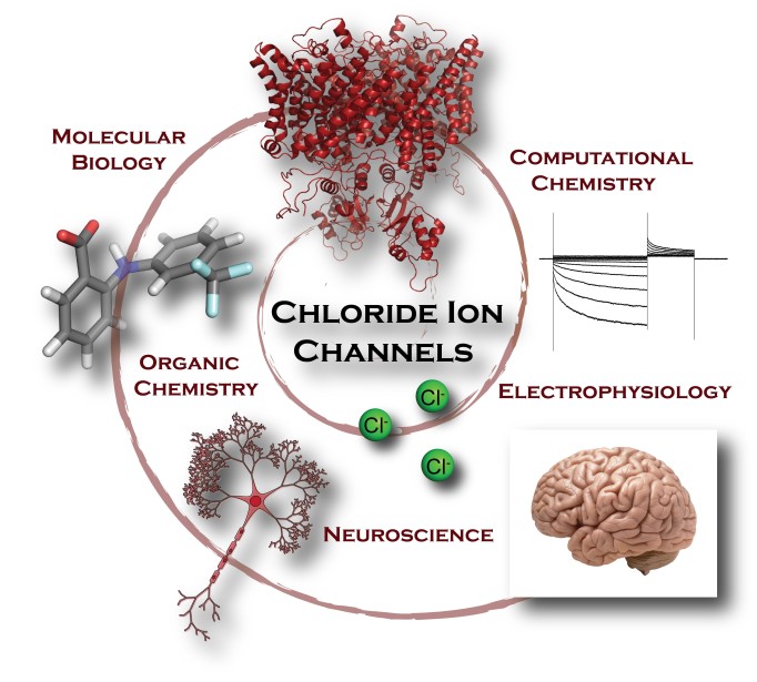
Scientists understand how these processes work at a basic level, but they involve an incredibly complex system of interactions, and there is a lot we still have to learn. My particular research project focuses on an understudied piece of this puzzle, a class of ion channels called CLC chloride channels. There are nine different subtypes of this protein in the human body, and generally speaking, they allow the passage of negative charge (in the form of chloride ions) across cell membranes. One of these, CLC-2, is highly expressed in both neurons and glia in the brain, but its function remains poorly understood. My research seeks to develop new molecular tools that will facilitate study of this ion channel both in the healthy brain and in disease states.

Why is your research important to the public?

The CLC-2 chloride channel is present in both neurons and glia in the brain. Neurons, which compose about 20% of the cells in the brain, are the electrically excitable cells that are responsible for the firing of action potentials. Glia, which are non-excitable cells, make up about 80% of your brain. People once thought these cells had only a passive structural function, but we now know that glia play a number of mechanistic roles in overall brain function and health. While we know that CLC-2 is present in the brain, much of the insight into CLC-2 function in this organ has come from selective gene knockout studies in mice. Mice lacking the CLC-2 gene exhibit severe white matter degeneration (leukoencephalopathy) over time, which suggests a critical role for CLC-2 in glial cells. In neurons, a variety of suggestions have been made regarding the role of CLC-2 in cellular excitability and ion homeostasis.

While mouse genetic studies provide clear evidence that CLC-2 has an important function in the brain, such studies are a rather crude approach to studying its specific function and behavior, particularly since genetic knockouts can cause compensatory changes in protein expression. To avoid such complications, an alternative approach is to use small-molecule tools that can rapidly and reversibly modulate CLC-2 function. For many ion channels, nature has provided such tools in the form of molecular toxins that organisms produce as a defense mechanism. One common example is tetrodotoxin (TTX), a potent neurotoxin that is found in pufferfish, ocean sunfish, and rough-skinned newts. The poison works by acting as a molecular cork in voltage-gated sodium channels, blocking the passage of sodium ions, which results in paralysis and stopping the heart of any potential predator. Chemists have isolated these naturally occurring toxins, determined their structures, and figured out how to synthesize these same molecules in the lab. In doing so, chemists are able to take advantage of the exquisite ability of these molecules to recognize and bind with high affinity to a single ion channel out of the thousands of types of proteins in the human body. In the lab, small-molecule tools like TTX allow scientists to study ion channels in a variety of ways. Such molecules can be used as structural probes of sodium channels expressed in cultured cells, or they can be used to evaluate how inhibition of sodium channels in brain tissue affects action potential firing and overall signal propagation. These molecular tools have been invaluable for studying how ion channels work at the biophysical and physiological levels.

In contrast to sodium channels, the number of small molecules that specifically recognize and bind to members of the CLC chloride channel family is extremely limited. Of the molecules that are known to bind and inhibit the flow of chloride current, most require high concentrations to have an effect and will non-specifically stick to a variety of other proteins in the body besides CLC channels. The challenge set before us as chemists was to design a novel molecular tool that would selectively and potently bind to CLC-2, in order to study its function in the brain. By employing a combination of structural biology, electrophysiology, modern computational methods, and chemical synthesis, we are developing new CLC-2 specific small-molecule leads for elucidating the role of CLC-2 in healthy brain tissue, as well as in disease states like epilepsy and leukoencephalopathy. This knowledge can provide the basis for understanding and treating a variety of neurological disorders.

BPS Brain Blog Graphic, Koster

How did you get into this area of research?

I came to graduate school at Stanford with a background in organic chemistry but with a strong interest in learning to apply these skills to tackle molecular problems in the biological sphere. With the School of Medicine directly across the street from the Department of Chemistry, it was easy to establish a collaboration between the Maduke and Du Bois labs to combine their expertise in electrophysiology and CLC ion channel physiology with chemical synthesis. This collaboration, together with fellowship and grant support from a number of interdisciplinary institutes at Stanford (Bio-X, the Center for Molecular Analysis and Design (CMAD), and the center for Chemistry, Engineering, and Medicine for Human Health (ChEM-H)), enabled us to take on this challenging project at the interface of chemistry and biology. We were particularly drawn to CLC-2 because of its fascinating yet poorly understood physiology in the brain and the lack of tools available to study it.

How long have you been working on it?

We have been working on this project for approximately 4 years.

Have you had any surprise findings thus far?

CLCs have traditionally been very challenging targets, and we had very few leads to follow when we began this project. At the outset, we were working with molecules with potency towards CLC-2 in the ~1 mM range and with little to no selectivity among the human CLC homologs (and even other classes of ion channels). Employing a relatively small automated patch-clamp electrophysiology screen, we identified a first lead compound with potency in the low-micromolar range. With further structural modification and rounds of structure-activity relationship studies, we improved the potency to mid-nanomolar levels. These newly developed molecules are the most potent small-molecule CLC inhibitors discovered to-date—it was a very exciting day in lab when we first found those results! We are currently working to understand the basis for molecular recognition between these small-molecules and the CLC protein with the goal of being able to rationally design better small-molecule tools that can act as tunable modulators (either activators or inhibitors) of CLC-2 function.

What is particularly interesting about the work from the perspective of other researchers?

We believe that our research will be particularly impactful for other researchers in the field of neuroscience. We have developed the most potent small-molecule CLC-2 inhibitor known to-date. This molecule can be used as a pharmacological tool to establish the physiological function of CLC-2 in the brain. In addition, we are working to modify the molecular scaffold of our CLC-2 inhibitor to provide tools with a variety of purposes. For example, by attaching biotin and fluorescent dyes, we aim to provide a tool for imaging and visualizing under a microscope exactly where these ion channels are located and how they are trafficked within neuronal and glial cells. Altogether, the molecular tools we are developing will be vital for studying the function of CLC-2 ion channels in tissue, cells, and at the atomic level.

What is particularly interesting about the work from the perspective of the public?

Given the significant advances in science and technology, it is remarkable how little we still know about the workings of the human brain. Most of the work involving electrical signaling in the brain has focused on sodium, potassium, calcium, and protons as carriers of charge, however it is clear that chloride transport plays a critical, yet understudied, role as well. In the 26 years since the discovery of the CLC-2 chloride channel, some progress has been made in understanding its role in the brain, but many questions remain, arguably due to the absence of selective tools for reversibly modulating CLC channel function in live cells.  It is our hope that the molecular tools we are developing will enable research to give us an enhanced understanding of CLC-2 in the brain and allow us to begin studying the possible role of CLC-2 in epilepsy and normal brain development. This work also has potential therapeutic impact in that the small molecules we are developing can offer prospective leads for drug development in the treatment of neurological disorders.

Using Biophysics to Understand Heart Disease

February is American Heart Month. Heart disease is the leading cause of death for men and women in the United States, causing 1 in 4 deaths each year. We spoke with Biophysical Society members Daniel Beard, University of Michigan, and Andrew McCulloch, University of California, San Diego, about each of their labs’ research related to heart disease.


MIP_Dan_Beard_0Daniel Beard
University of Michigan

What is the connection between your research and heart disease? Why is your research important to those concerned about heart disease?

A major focus of our research is on the link between cardiac function and myocardial metabolism. In physical exercise, as the rate of work done by the heart increases compared to rest, the rate of cardiac ATP hydrolysis increases commensurately. By building computer models to represent kinetic control of oxidative (mitochondrial) ATP synthesis, we have been able to build a robust framework for simulating ATP supply/demand matching in vivo [1, 2]. Once we had a working understanding of physiological regulation of ATP synthesis in the heart, we used that working knowledge as a starting place to explore how the physiological system becomes dysfunction in heart disease. In particular, it has long been recognized that in heart failure—a condition in which the mechanical pumping ability of the heart is compromised—concentrations of adenosine triphosphate (ATP), its hydrolysis products, and related metabolites are depleted compared to normal. Neither the causes nor the consequences of these changes are well understood. We think that one consequence of impaired energy metabolism is that it directly contributes to impaired mechanical function. Our current research is squarely focused on determining these causes and consequences of mechano-energetic dysfunction, and on finding new ways to repair/restore myocardial metabolism to improve mechanical function in heart failure.

How did you get into this area of research?

My interest in this area goes back to work that was part of my PhD thesis, working in Jim Bassingthwaighte’s lab on simulating physiological transport of oxygen, and other solutes. I became increasingly interested in oxygen transport, and questions of how oxygen delivery is matched to oxygen demand in the myocardium. In this context I started to look in some depth at the physiological regulation of oxidative phosphorylation, not for its own sake, but because I was interested in how it contributed to governing oxygen transport. In other words, at first mitochondrial metabolism was on the periphery of what I was initially interested in. That peripheral interest gradually grew into a major research thrust.

Do you receive public funding for this work? If so, from what agency?

Yes, we have been fortunate to be supported for this work, primarily from the National Institutes of Health National Heart Lung and Blood Institute.

Have you had any surprise findings thus far? 

Our first big surprise followed from my lab’s first NIH funding award, for which we proposed to investigate a conundrum in the field cardiac energy metabolism: How is ATP synthesis supply matched to changing demand levels in light of apparently constant concentrations of ADP and inorganic phosphate (Pi) in the heart? Our answer to this question is that, in fact, the ATP supply is not matched to demand while maintaining constant levels of ADP and Pi. Specifically, our model-based analysis of the in vivo data on phosphate metabolite levels in the heart predicts that changing [Pi] provides a critical feedback signal to stimulate increasing ATP synthesis with increasing rates of ATP hydrolysis. This simple idea fundamentally challenges established ideas in the field. In fact, our hypothesized role of Pi as a primary feedback signal for oxidative ATP synthesis in the heart is still at least a little bit controversial and it would be incorrect to say that it is universally accepted. Regardless, the hypothesis has survived all of our attempts to disprove it!

Beard Image

The next big surprise came when we applied our models of cardiac energy metabolism to analyze data from animal models of heart failure. We found that hallmarks of myocardial energetics in heart failure—diminished ATP and ATP hydrolysis potential—could be effectively captured by simulations in which mitochondrial function is normal. At first blush that finding seems contradictory: Since 95% of ATP in the heart is produced by mitochondria, how can diseased hearts with diminished ATP have normally functioning mitochondria? The explanation pointed to by our analysis is that reduction in cytoplasmic metabolic pools is a critical driver of energetic/metabolic dysfunction in the failing heart. Those predictions were later put to the test when, in collaboration with Igor Efimov’s lab, we were able to measure mitochondrial function in samples from failing  human hearts, revealing no significant dysfunction compared to healthy controls [3]. This is not to say that mitochondrial function is normal in the heart in heart failure, but rather that we believe dysfunction in mitochondrial energy metabolism is not necessarily intrinsic to the mitochondria themselves, but rather driven by the local environment they find themselves in.

What is particularly interesting about the work from the perspective of the public?

Patients with heart failure wind up seeing a cardiologist, not with complaints of impaired cardiac energetics, but with health problems—shortness of breath, exercise intolerance—directly related to impaired heart pumping. Yet we think that metabolic/energetic dysfunction can contribute directly to mechanical dysfunction in heart failure. Our current research in this area is on the link between mechanics and energetics. We are using computer models that integrate metabolic and mechanical function to better understand the physiological connection between chemical and mechanical function in the heart, to determine ways in which this connection breaks down in heart disease, and identify new strategies to improve mechanical pump function by restoring the metabolic state.

  1. Wu, F., et al., Phosphate metabolite concentrations and ATP hydrolysis potential in normal and ischaemic hearts. J Physiol, 2008. 586(17): p. 4193-208.
  2. Bazil, J.N., D.A. Beard, and K.C. Vinnakota, Catalytic Coupling of Oxidative Phosphorylation, ATP Demand, and Reactive Oxygen Species Generation. Biophys J, 2016. 110(4): p. 962-71.
  3. Holzem, K.M., et al., Mitochondrial structure and function are not different between nonfailing donor and end-stage failing human hearts. FASEB J, 2016.

Andrew3Andrew McCulloch
University of California, San Diego

What is the connection between your research and heart disease?

We use in-vitro and in-vivo experiments primarily using mouse models of heart failure and arrhythmias together with multi-scale computational models to discover cellular and molecular mechanisms of electrical and mechanical dysfunction at the tissue and organ scales.

Why is your research important to those concerned about heart disease?

Our research is becoming of particular interest to cardiologists and patients with heart diseases because we have started to discover ways to apply the computational modeling tools we validated in the lab to analyze and predict therapeutic outcomes in patients, including heart failure patients with electrical dyssynchrony who are indicated for cardiac resynchronization therapy, patients with atrial fibrillation or at risk of ventricular fibrillation who can benefit from radiofrequency ablation therapy, and children and adults with congenital heart diseases who are at risk of developing heart failure or arrhythmias later in life.

How did you get into this area of research?

It started as a MS thesis project in Engineering Science when I was 19, that turned into a PhD in Physiology and Engineering and then a faculty career.

How long have you been working on it?

Over 35 years.

Do you receive public funding for this work? If so, from what agency?

Yes, we rely almost exclusively from NIH funding via the NHLBI, NIBIB and NIGMS though we have also received valuable support in the past from the NSF, NASA, DARPA, AHA and Heart Rhythm Society.

image1.001

Have you had any surprise findings thus far?

Yes, we have found with and novel mouse and computational models that phosphorylation of specific serine residues on cardiac myosin regulatory light chain not only affect crossbridge dynamics, but also give rise to a feedback that affects the calcium-dependent active of the thin filament. We have also found that patient-specific models have the unexpected potential to predict and optimize the outcomes of device therapies for heart failure.

What is particularly interesting about the work from the perspective of other researchers?

The power of modern multi-scale and systems biology modeling to help understand genotype-phenotype relations in animal models of heart diseases.

What is particularly interesting about the work from the perspective of the public?

The potential for computer models to improve the early diagnosis and clinical management of heart diseases in adults and children.

Biophysics on World AIDS Day

December 1 is World AIDS Day. Globally, there are an estimated 36.7 million people who have the virus. Despite the virus only being identified in 1984, more than 35 million people have died of HIV or AIDS, making it one of the most destructive pandemics in history.

In recognition of World AIDS Day, we spoke with Biophysical Society member Leor Weinberger, University of California, San Francisco, whose research focuses on how HIV makes a fate decision between active replication and latency.

LSW image

What is the connection between your research and HIV/AIDS?

Our lab focuses on fundamental aspects of how HIV makes a fate decision between active replication (turning on) and a long-lived dormant state called latency (turning off).  Latency is the chief barrier to curing a patient of HIV.  We use quantitative single-cell approaches and mathematical models to map the transcriptional circuitry that controls this fate decision.

Many years ago we found that HIV harnesses and amplifies stochastic fluctuations (noise) in gene expression to control its active vs. latent fate decision (back in the early 2000’s HIV, in fact, provided the first experimental evidence of a noise-driven fate decisions).  Recently, we discovered that noise can be manipulated with small molecules and we are developing therapies that disrupt HIV circuitry by manipulating noise.

Why is your research important to those concerned about HIV/AIDS?

HIV latency is the chief barrier to curing a patient of HIV.  There is no vaccine for HIV and current anti-retroviral therapies only halt active viral replication; if a patient discontinues therapy, HIV will reactivate from latently infected cells and viral levels in the blood rebound to pre-treatment levels within a few weeks.  We now know the most problematic (i.e., longest-lived) latent reservoir exists in a type of white blood cell called a CD4+ T cell, the same cell type that HIV actively infects.  HIV remains ‘silent’ and integrated as a “provirus” in the genome of these cells.  So, the field is actively pursuing approaches to better understand and attack the proviral latent reservoir.

Our work showed that a major mechanism HIV uses to promote its silencing is harnessing stochastic fluctuations in a transcriptional feedback circuit and gene-expression noise is now acknowledged as a primary clinical barrier to reversing HIV latency and curing HIV. Recently, we identified molecules that modulate stochastic fluctuations and could be a new class of therapeutic candidates for reversing latency.

How did you get into this area of research and how long have you been working on it?

I’d always hoped to make a contribution to medicine but as a student I became enchanted with the beauty of nonlinear dynamics theory and the intellectual approach of physics.  This eventually led me to train with a group of physicists who were developing mathematical models to explain HIV infection dynamics in patients.  At the time many physicists were modeling biology but very few models were grounded by experiment (HIV was one of the very few).  A problem, however, was convincing experimental collaborators to test the most interesting predictions.  So, in grad school I decided to learn wet-lab molecular biology and eventually started my own experimental lab.

This combination of modeling and experiment helps the science move faster.  Plus, I greatly enjoy training students and postdocs at the interface of these disciplines and translating basic discoveries into potential therapies is both fun and rewarding.  So, my lesson to aspiring biophysicists is: try to find training opportunities that cross disciplinary interfaces (theory and experiment, basic and applied, etc…) it can be challenging at times but is also very rewarding.

Do you receive public funding for this work? If so, from what agency?

We received generous funding to develop a radical new class of ‘autonomous’ antiviral therapies from the Defense Advanced Research Projects Agency (DARPA) as well as the NIH, especially the NIH Director’s Common Fund.  DARPA actually started a program called INTERfering and Co-Evolving Prevention and Therapy (INTERCEPT) to fund this work.  The NIH Common Fund was what allowed us to start out with this research; it is a unique and important program that funds high-risk, high-reward research through the Director’s Pioneer and New Innovator Awards and has mechanisms to support young researchers setting off in bold directions.

Have you had any surprise findings thus far?

Probably our most surprising finding was discovering noise-enhancer molecules (Dar et al. Science 2014).  These are small molecules (some are approved drugs) that alter transcriptional fluctuations without changing the mean level of gene expression and act like Bunsen burners for cells: they appear to potentiate cell-fate decisions in diverse systems.

The analogy can be made to physical and chemical systems, where many processes are enhanced by catalysts but also by increasing the thermal fluctuations (e.g., kT in the Arrhenius equation).  Catalysts deterministically lower activation-energy barriers on potential-energy landscapes but when deterministic drivers are insufficient to cross the barrier, amplifying thermal fluctuations (e.g., with a Bunsen burner) provides an added perturbation for crossing activation-energy barriers.

Remarkably, we found that this concept applies to gene regulation during cell-fate decisions; the novel class of noise-enhancer molecules act like biological Bunsen burners.  As a model system, we focused on HIV where the leading HIV-cure strategy requires latent virus be reactivated and then purged—but where current reactivation schemes are ineffective.  Strikingly, these Bunsen burner-like noise-enhancer compounds potentiated transcriptional activators to greatly enhance HIV reactivation in patient cells.  We also also identified noise-suppressor compounds (effectively ‘ice packs’) that inhibit reactivation.

It is important to note that noise modulators starkly contrast with generic stress responses (e.g., starvation), which can increase noise but necessarily attenuate transcriptional activators.  Thus, noise modulation is a fundamentally departure from generic stress responses.  Preliminary evidence indicates that noise-modulating molecules could provide a general tool to manipulate diverse cell-fate decisions from antibiotic persistence to cellular reprogramming and cancer.

What is particularly interesting about the work from the perspective of other researchers?

I often hear from colleagues that they most appreciate that our work shows a biological role for stochastic ‘noise’.  Before the HIV example, there had been beautiful and elegant work from colleagues measuring and identifying the sources of stochastic gene-expression noise in laboratory organisms (E coli and yeast).  However, it was not clear if cells ‘cared’ about the noise or simply ignored it.  The HIV example (Weinberger et al. Cell, 2005) was the first to experimental evidence that stochastic noise in gene-expression could flip a genetic switch and drive a biological fate decision (i.e., developmental bet-hedging).  Developmental bet-hedging–the concept that organisms harness intrinsic variability to enable bet-hedging decisions between alternate developmental fates, similar to how financial houses diversify assets to minimize risk in volatile markets—had in fact been hypothesized since the 1960s.  Subsequent work from some of my scientific heroes—and now friends—showed similar results in B subtillis, stem cells, and cancer cells.  Recently, we have gotten a lot of requests from colleagues for our noise enhancer molecules and we send out samples every few weeks.  I suspect that these molecules may end up being the more helpful contribution to the field.

What is particularly interesting about the work from the perspective of the public?

Typically, it is a different thread of research in our lab that catches the public’s attenuation.  Many years ago we hypothesized the idea of treating infectious diseases by engineering therapies that co-evolve and co-evolve TIPs mentioned above (Metzger et al. PLoS Comp. Biol. 2011) has garnered the most public interest.  Existing measures for infectious disease control face three ‘universal’ barriers: (i) Deployment (e.g. reaching the highest-risk, infectious ‘superspreaders’ who drive disease circulation); (ii) Pathogen persistence & behavioral barriers (e.g. adherence); (iii) Evolution (e.g. resistance and escape). To surmount these barriers, we have proposed a radical shift in therapeutic paradigm toward developing adaptive, dynamic therapies (Metzger et al. 2011). Building off data-driven epidemiological models, we show that engineered molecular parasites, designed to piggyback on HIV-1, could circumvent each barrier and dramatically lower HIV/AIDS in sub-Saharan Africa as compared to established interventions. These molecular parasites essentially steal replication and packaging resources from HIV within infected cells thereby generating Therapeutic Interfering Particles (TIPs), which deprive HIV of critical replication machinery thereby reducing viremia. The fundamental departure from conventional therapies is that TIPs are under strong evolutionary selection to maintain parasitism with HIV and will thus co-evolve with HIV, establishing a co-evolutionary ‘arms race’ (Rouzine and Weinberger, 2013).  Like Oral Polio Vaccine, (OPV)—currently used for the W.H.O. worldwide polio-eradication effort—TIPs could also transmit between individuals, a recognized benefit for OPV.  TIP transmission would occur along HIV-transmission routes (via identical risk factors), thereby overcoming behavioral issues and automatically reaching high-risk populations to limit HIV transmission even in resource-poor settings.

Importance of Biophysics in Breast Cancer Progression

October is Breast Cancer Awareness Month in the US. We spoke with University of California, San Diego graduate student Pranjali Beri and her PI, BPS member Adam J. Engler, about their research on breast cancer and other epithelial-based cancers. 


What is the connection between your research and cancer?

Cancer is the second leading cause of death in the US, resulting in approximately 600,000 deaths in 2016. The negative prognosis associated with cancer is due in large part to metastasis of a primary tumor. Cancer metastasis is the process by which tumor cells leave the primary tumor, enter the blood stream (intravasation), exit the blood stream at a different site in the body (extravasation), and establish a secondary tumor. However, tumors are exceedingly heterogeneous and only a small fraction of cancer cells from the primary tumor are capable of establishing secondary tumors. The metastatic potential of identical solid tumor types also varies from patient-to-patient due to expression differences of critical markers, making it nearly impossible to identify a universal biomarker that can predict metastatic potential of all solid tumors.

fig 1 A

Cancer cell migration away from the primary tumor is driven, in part, by physical interactions between cells and the surrounding extracellular matrix. Protein clusters known as focal adhesions allow cells to attach to the matrix proteins, and stability and strength of these attachments plays a role in regulating cancer cell migration. Our research is attempting to understand the link between adhesion strength via focal adhesions and cancer cell dissemination. In our recently publication, we quantify population adhesion strength of various epithelial cancer cell lines by utilizing a spinning-disk shear assay (Figure 1a). The shear stress required to detach 75% of the cell population serves as a metric to describe the adhesion strength of that population. In the presence of conditions that mimic the tissue adjacent to tumors, e.g. low divalent cations, we found that heterogeneous adhesion strength for the most aggressive cells indicate that subpopulations within aggressive cell lines were capable of metastatic behavior. This is similar to the small fraction of the primary tumor previously thought to contain stem cell-like properties of self-renewal, differentiation, and migration.

fig 1 B

Currently, our research further seeks to sort, capture, and analyze cells with more labile focal adhesions in response to stromal cation concentrations. We have developed a parallel plate flow chamber assay to isolate weakly adherent cells (Figure 1b) and characterize their migratory propensity in relation to strongly adherent as well as unselected cell populations. By demonstrating that there is a link between adhesion strength and migratory propensity of the cancer cells, we can use it as a biophysical marker for metastatic potential.

Why is your research important to those concerned about cancer?

Epithelial tumors, or carcinomas, are the most common type of cancer. There is no universal biomarker that acts as an indicator for metastatic potential. However, most epithelial cancers undergo metastasis. Having a physical indicator of metastasis can be beneficial in identifying the aggressiveness of a tumor and its likelihood of forming secondary metastases, independent of the type of epithelial tumor that it is.

How did you get into this research?

Throughout my undergraduate career, I have been interested in microfluidic devices and their applications as diagnostic devices. In graduate school, I joined the Engler lab in order to apply my microfluidics background towards cancer metastasis research.

How long have you been working on it?

I began working with microfluidic devices during my undergraduate studies. However, it was in graduate school that I used it to study cancer cell dissemination.

Do you receive public funding for this work? If so, from what agency?

I am currently funded by the National Science Foundation through their Graduate Research Fellowship Program. This research is also funded by the National Institute of Health and the Department of Defense Congressionally Directed Medical Research Program.

Have you had any surprising findings thus far?

Tissue adjacent to tumors has dramatically lower ion concentrations than in the tumor. In our previous publication, heterogeneity is most pronounced in highly metastatic cancer cell lines, but only when exposed to low ion conditions that mimic adjacent tissue; in the presence of high cation concentration, akin to the tumor microenvironment, metastatic cells are mechanically indistinguishable from their non-metastatic counterparts. This shift in adhesion strength was not present in non-metastatic cancer cell lines but was present in epithelial cancer cell lines from other tumors as well, including prostate and lung. While these previous studies could isolate the strongly adherent fraction remaining attached, recent experiments using flow chamber assays indicate that weakly adherent cells from the same cell lines display increased migration speed and are more processive in comparison to unsorted or strongly adherent populations. These results indicate that adhesion strength can potentially act as a biophysical marker of metastatic potential, and that the weakly adherent cells are likely to have the highest metastatic potential.

What is particularly interesting about this work from the perspective of other researchers?

Our fluidic-based separation method could allow us to isolate cancer cells by their metastatic potential. The adhesion strength-based separation method can serve as a potential prognostic device that exposes patient biopsies to shear stress, correlates weakly adherent cell isolation with metastatic potential, and makes a prognostic determination about the likelihood to metastatic disease in the future.

What is particularly interesting about this work from the perspective of the public?

By establishing the link between adhesion strength of the cells in the tumor and the metastatic potential of the tumor, we can ascertain the aggressiveness of patient tumors and tailor treatments accordingly.

Understanding Alzheimer’s Disease through Biophysics Research

September 21 is World Alzheimer’s Day. Alzheimer’s disease is the leading cause of dementia, from which 47 million people worldwide suffer. It affects memory, thinking, orientation, comprehension, calculation, learning capacity, language, and judgement. 

In recognition of World Alzheimer’s Day, we spoke with two Biophysical Society members whose research aims to improve understanding of the mechanisms behind Alzheimer’s and other neurodegenerative diseases.


Liz Rhoades, University of Pennsylvania

Teele10-18vtyq3-200x300

What is the connection between your research and Alzheimer’s disease?

We study the protein tau, which is a microtubule associated protein.  In Alzheimer’s and other neurodegenerative disorders, tau forms fibrillar aggregates that are deposited in brain tissue.  There are six isoforms of tau found in adult humans and alteration in the amounts of the isoforms are linked to disease development. Interactions of tau with microtubules are normally regulated by phosphorylation and tau aggregates derived from patient tissues are hyperphosphorylated, providing a link between loss of native tau function and disease as well.

Why is your research important to those concerned about Alzheimer’s?

We are working to understand basic aspects of tau function because we of the insight it provides to loss of function in disease.  Despite rather intensive study, molecular details of tau function are still lacking.  This is at least in part due to the fact that tau is large intrinsically disordered protein and thus it is challenging to characterize its structural features, particularly when associated with tubulin (the soluble building blocks of microtubules) and microtubules. For example, a few years ago, we observed that tau binds to soluble tubulin, a feature that had not previously received much attention. Our results suggests that it binds with similar affinity to tubulin as it does to microtubules which suggests that understanding how mutation impacts its interactions with tubulin is as important as characterizing how It interacts with microtubules. This is important because therapeutic strategies may very well involve targeting interactions between tau and its functional binding partners – we need to know who those partners are and the relevant features of the interaction!

Rhoades

The image here is from a paper that was published in PNAS last fall. It shows tau binding to two soluble tubulin dimers, and is based on our single molecule FRET measurements. It highlights how tau retains a primarily disordered state while binding and initiating tubulin polymerization. 

How did you get into this area of research?

We had been looking at tau aggregation in the lab for a couple of years, and then I had a few students – two graduate students and an undergraduate –  who were very interested in working with tubulin.  They were they ones who really pushed to get things up and running.  Anyone who has ever done a tubulin purification in their lab knows that this is not a trivial undertaking!

How long have you been working on it?

6 or 7 years

Do you receive public funding for this work? If so, from what agency?

In the past we received funding from NSF (MCB)  and currently we receive funding from NIH (NIA).

Have you had any surprise findings thus far?

I think most findings I get excited about are surprises, but there are three that I can think of as particularly surprising.  The first was that tau binding to soluble tubulin had been largely overlooked in previous studies. We started working with soluble tubulin because it was easier for us to use in single molecule experiments (one of our primary tools) and only as we began to get interested results, do we recognize that there really was not a deep literature on tau-tubulin. The second was the tau point mutants linked to different neurodegenerative disorders bound more tightly to tubulin than the wild-type tau.  Our expectation based on the tau-microtubule literature was the mutation should decrease the binding affinity.  We are still working to understand the impact of this on tau function.  The third is that a region which flank the microtubule binding region has a high affinity for tubulin and allows for tau to bind to multiple tubulin dimers simultaneous to form a ‘fuzzy complex’.  This work was published in Biophysical Journal this summer.

What is particularly interesting about the work from the perspective of other researchers?

I think some of it is probably methodology – we are using single molecule FRET and FCS to investigate tau-tubulin and working to make useful measurements in relatively complex, heterogeneous systems.

What is particularly interesting about the work from the perspective of the public?

Understanding tau’s interactions with native binding partners may provide new targets for therapeutics.  I think anyone who has a family member or loved one who suffers from Alzheimer’s or another neurodegenerative disorder would find that interesting.


Dieter Willbold, Heinrich-Heine-Universität Düsseldorf and Forschungszentrum Jülich

image description

What is the connection between your research and Alzheimer’s disease?

My interest is focused on three-dimensional structures and dynamics of medically relevant proteins at atomic resolution and their interactions with native and artificial ligands. Autophagy and neurodegenerative diseases, which by the way do have a clear connection with each other, fall within my main interest areas. I want to understand protein aggregation in time and space at high resolution. And, I want to develop strategies and compounds that allow intervention and prevention. And the protein I am working on now for a very long time as a researcher is the Alzheimer’s disease (AD) related amyloid-beta protein (Aβ).

Why is your research important to those concerned about Alzheimer’s?

We do a lot of in-depth basic science on aggregation of Aβ, tau, alpha-synuclein, and many other proteins. We also design compounds for novel disease intervention strategies, and develop novel biomarkers and assays to measure them appropriately and most sensitively. All three, basic science, drug design and biomarker development, are based on biophysical principles and physico-chemical “thinking” and heavily rely on respective methods, such as NMR, x-ray crystallography, cryo-electron microscopy, ultracentrifugation, surface plasmon resonance, micro-calorimetry, TIRF microscopy, AFM, micro-thermophoresis, biolayer interference, and all kinds of spectroscopies.

Willbold

Figure 1: Cross section through the Aβ fibril illustrating the stepwise overlapping arrangement of the Aβ proteins. (Copyright: Forschungszentrum Jülich / HHU Düsseldorf / Gunnar Schröder). See also: http://www.fz-juelich.de/SharedDocs/Pressemitteilungen/UK/EN/2017/17-09-08-alzheimer-fibrillen.html .

How did you get into this area of research?

Already during my PhD project, which was mainly on the 3D structure determination of the transactivator protein (Tat) of the equine homologue of the HIV virus, I was engaged in structural studies of the amyloid-beta protein (Aβ) by NMR spectroscopy with some of the results published in 1995 with Paul Rösch being my supervisor and mentor. Ever since then, I was thinking of potential therapeutic intervention strategies. Since 1999, when I was heading my own junior research group in Jena, I had the necessary resources to at least start research on intervention strategies.

Soon after, I became involved in projects on prion diseases and prion protein (PrP) aggregation, when I accepted my first professorship at the Heinrich Heine University Düsseldorf in very close collaboration with Detlev Riesner. The common themes in these protein misfolding or protein aggregation diseases became quite clear. In my view, any intervention strategy – rather than a prevention strategy – needed to target toxic aggregates and get rid of them, rather than to reduce the formation of the monomeric species. As a biophysicist, I thought it would be a good idea to shift equilibria between monomers and aggregates away from the toxic aggregates (or oligomers as they are called today). To do this, we looked for compounds that bind to Aβ monomers with the free binding energy being used to lower the free energy level of monomers thus shifting the thermodynamic equilibrium towards Aβ monomers. The wording we use nowadays is that such a compound stabilizes Aβ in an aggregation-incompetent conformation. Because this is also happening with Aβ monomer units within Aβ oligomers, such a compound is also able to damage and destroy already pre-formed Aβ oligomers leading ultimately to their elimination. To identify a useful lead compound, we used mirror image phage display selection, a tool that allows selection of a compound from huge peptide libraries, but yielding a fully D-enantiomeric peptide, that does not have the disadvantages of normal L-peptides, which are very easily degraded and immunogenic. Our lead compound with the name “D3” (D-peptide from the third selection trial) showed really nice properties in vitro and in vivo. When we then wanted to elucidate the mechanism of action, it was essential to establish a whole zoo of methods and assays, which brought me even deeper into the field of protein aggregation in general and Alzheimer’s in particular. I just wanted to elucidate and pinpoint the mechanism of action and to reveal structural details of any interaction of Aβ with itself and with ligands.

Do you receive public funding for this work? If so, from what agency?

Yes, now we do. In the beginning, I was not successful at securing additional money from funding agencies, e.g., the DFG. The project received great review reports about the underlying idea, but the panels ultimately decided that the project was too risky. Therefore, I used most of the institutional resources (which was not much in those years) for the project. Only in 2007, the Volkswagen-Stiftung funded a side project. Since 2013, I did and still do receive significant funding from the Helmholtz-Gemeinschaft, the federal ministry BMBF, the EU, and also from the Michael J Fox Foundation, the Weston Brain Institute, Alzheimer’s Research UK, as well as the Alzheimer’s Association. We have also been part of a JPND network.

Have you had any surprise findings thus far?

Yes indeed — many! Just to describe some: our lead compound D3 worked effectively not only in vitro, but also in several animal models. This has been a successful long-term collaboration with my dear colleagues Thomas van Groen, Inga Kadish and Antje Willuweit. D3 improved cognition in several models and decelerated neurodegeneration in an additional animal model that we have received from my dear collaborator Uli Demuth. We established an assay called QIAD that allows us to quantify Aβ oligomer elimination efficiency (https://www.ncbi.nlm.nih.gov/pubmed/26394756). We found that D3 efficiently eliminates Aβ oligomers, but many compounds that have already been in the clinics and failed are not able to do this. By following aggregation of N-terminally truncated and pyro-glutamate-modified Aβ (pEAβ) by NMR and CD spectroscopy, we found intermediates with helical secondary structure during aggregation.

When we tried to follow Aβ aggregation by SANS and analytical ultracentrifugation (AUC), we did not find any intermediates between monomers and penta- or hexamers (https://www.ncbi.nlm.nih.gov/pubmed/28559586). Thus, Aβ seed formation may be a reaction of very high order. In our recent research, (7th Sep 2017, https://www.ncbi.nlm.nih.gov/pubmed/28882996) we published a high resolution cryo-EM structure of Aβ fibrils. This structure provided many surprising findings in one hit including: all 42 residues of Aβ(1-42) are part of the fibril structure, there is no C2 symmetry between the two proto-filaments of the fibril, both ends of the fibril are different, each Aβ monomer subunit contacts many other subunits and six Aβ monomers form the minimal fibril unit. See also the respective report in alzforum.org: http://www.alzforum.org/news/research-news/amyloid-v-fibril-structure-bares-all .

What is particularly interesting about the work from the perspective of the public?

I think that from the perspective of the public, two questions are relevant and interesting: Can we visualize highly complex things? Especially as structural biologists, we indeed can. Just look at the beautiful picture of the Aβ fibril at atomic resolution below. The second question is, of course: Can we contribute to efforts for improving the quality of life, for example by developing therapeutic strategies and drug candidates? Yes, we can also do (or at least try to do) this. It is, however, a huge undertaking that needs substantial funding and teamwork with many experts and specialists that you would not contact for basic science.

Just today (18th Sep 2017), we founded a company named Priavoid that will take an optimized derivative of D3 into clinical studies and hopefully to the market someday. In parallel, because Aβ oligomer elimination is our most favored mechanism of action, we have developed a technology called sFIDA (surface-based fluorescence intensity distribution analysis), which is able to quantify Aβ oligomers in body liquids like CSF and blood at single particle sensitivity (https://www.ncbi.nlm.nih.gov/pubmed/27823959). The development of this technology was and is important to ultimately show target engagement of our Aβ oligomer eliminating compounds. sFIDA will also be useful for early diagnosis of any protein misfolding disease, to recruit the “right” patients for clinical studies and to follow treatment success, if there is one. Thus, all in all, I think we have developed interesting results for the public domain.

Is there anything else you would like to add?

During the initial stages of the above described project to develop a novel therapeutic strategy for AD and to identify suitable compounds, there was only myself and my PhD student, Katja Wiesehan. Currently, there are many colleagues and coworkers that are the most experienced experts in their fields. It is such a beautiful experience to work and think with all of them and all the junior researchers, and to finally get things done. Please, have look at them and their groups in Düsseldorf (http://www.ipb.hhu.de/en.html) and Jülich (http://www.fz-juelich.de/ics/ics-6/EN/) with outstations in Grenoble and Hamburg. Finally, I shall not forget the many, many collaborators, of which I named only a few above.

Highlighting Biophysics Research During Sickle Cell Awareness Month

September is National Sickle Cell Awareness Month in the United States. Sickle cell disease is an inherited blood disorder that affects approximately 100,000 Americans and millions worldwide. It is particularly common among people whose ancestors come from Sub-Saharan Africa, South America, Cuba, Central America, Saudi Arabia, India, and Mediterranean countries such as Turkey, Greece, and Italy.

To recognize the awareness month, we spoke with BPS member George Em Karniadakis, Brown University, and his collaborators Xuejin Li, Brown University, and Ming Dao, MIT, about their research related to sickle cell disease. Their research was also featured on the cover of the July 11, 2017, issue of Biophysical Journal.


BPJ_113_1.c1.indd

What is the connection between your research and sickle cell disease?

Sickle cell disease (SCD) is the first identified molecular disease affecting more than 270,000 new patients each year. Our interests are in modeling multiscale biological systems using new mathematical and computational tools that we develop in our teams at Brown University and MIT in conjunction with carefully selected microfluidic experiments at MIT. We have an ongoing NIH-funded joint project that focuses on developing such validated predictive models for the sickle cell disease (SCD). In this project, with close collaboration between clinicians, experimentalists, applied mathematicians and physical chemists, we have been  developing new predictive patient-specific models of SCD, linking sub-cellular, cellular, and vessel-level phenomena spanning across four orders of magnitude in spatio-temporal scales. So far we have developed a validated patient-specific and data-driven multiscale modeling approach to probe the biophysical mechanisms involved in SCD from hemoglobin polymerization to vaso-occlusion.

Why is your research important to those concerned about sickle cell disease?

SCD is one of the most common genetic blood disorders that can cause several types of chronic and acute complications such as vaso-occlusive crises (VOC), hemolytic anemia, and sequestration crisis. It is also the first identified molecular disease (as early as 1947 by Linus Pauling), and the underlying molecular cause of the disease has been understood for more than half a century. However, progress in developing treatments to prevent painful VOC and associated symptoms has been slow. Consequently, we have been developing a “first-principles” multiscale approach that can handle the disparity of molecular, mesoscopic and macroscopic phenomena involved in SCD simultaneously. Such simulations could potentially answer questions concerning the links among sickle hemoglobin (HbS) polymerization, cell sickling, blood flow alteration, and eventually VOC. We hope, in turn, that these models will help in assessing effective drug strategies to combat the clinical symptoms of this genetic blood disorder.

Karniadakis

Figure 1. Dynamic behavior of individual sickle RBCs flowing in microfluidic channel. Inside the yellow circles are trapped sickle RBCs at the microgates, and inside the white circles are deformable RBCs, which are capable of circumnavigating trapped cells ahead of them by choosing a serpentine path (indicated by the white arrows).

How did you get into this area of research?

We have been working on multiscale modeling of blood disorders for more than 10 years.  In the very beginning, we were interested in developing new computational paradigms in multiscale simulations, which would enable us to perform multiscale realistic simulation of blood flow in the brain of a patient with an aneurysm. We then realized that the mesoscopic modeling of red blood cells (RBCs) and hemorheology in general seems to be the most effective approach for modeling malaria and other hematologic disorders. Then, we shifted our attention to the particle-based modeling of blood flow by employing the dissipative particle dynamics (DPD) method, which can seamlessly represent the RBC membrane, cytoskeleton, cytosol, surrounding plasma, and even the parasite in the malaria-infected RBCs. We developed multiscale RBC models and employed them to predict mechanical and rheological properties of RBCs and quantify molecular-level mechanical forces involved in bilayer–cytoskeletal dissociation in blood disorder. In 2012, we started to work on SCD, after realizing that no multiscale simulation studies of SCD had been conducted before – our work is the first of its kind!

How long have you been working on it?

As we mentioned above, we have been working in this field for more than 10 years.

Do you receive public funding for this work? If so, from what agency?

Yes, we receive support from NHLBI, the institute within NIH focusing on blood disorders based on the interagency funding initiative pioneered by Dr. Grace Peng. For those who are interested in this multiscale consortium they can visit: https://www.imagwiki.nibib.nih.gov/

Have you had any surprise findings thus far?

Plenty! For example, at the vessel scale, using computer models, we have discovered that it is the soft and sticky type of RBCs that initiate the blockage process and lead to sickle cell crises and not the rigid sickle cells! This is the first study to identify a specific biophysical mechanism through which vaso-occlusion takes place. At the cellular scale, we have developed a tiny microfluidic device that can analyze the behavior of blood from SCD patients. Informed from the microfluidic experiments conducted by Dr. Ming Dao’s group at MIT, we have developed a unique patient-specific predictive model of sickle RBCs to characterize the complex behavior of sickle RBCs in narrow capillary-like microenvironment. At the sub-cellular (molecular) scale, we have developed a particle HbS model for studying the growth dynamics of polymer fibers (recent cover of Biophysical Journal). The simulations provide new details of how SCD manifests inside RBCs, which could help other medical researchers in developing new treatments.

What is particularly interesting about the work from the perspective of other researchers?

It is known that the primary cause of the clinical phenotype of SCD is the intracellular polymerization of sickle hemoglobin (HbS) resulting in sickling of RBCs in deoxygenated conditions. However, the clinical expression of SCD is heterogeneous, making it hard to predict the risk of VOC, and resulting in a serious challenge for disease management. Our data-driven stochastic multiscale models, based on particle methods, can be used to explore and understand the dynamics of collective processes associated with vaso-occlusion that links together sub-cellular, cellular, and vessel phenomena. A similar computational framework can be applied to study blood flow in other hematologic disorders, including malaria, hereditary spherocytosis and elliptocytosis, as well as other blood pathological conditions in patients with diabetes mellitus or AIDS.  For example, in ongoing work we have quantified the biophysical characteristics of RBCs in type-2 diabetes mellitus (T2DM), from which the simulation results and their comparison with currently available experimental data are helpful in identifying a specific parametric model that best describes the main hallmarks of T2DM RBCs.  Perhaps, the most important extension is to connect such multiscale models to all the “omics” technologies (genomics, proteomics, metabolomics, etc.) to implement the vision of precision medicine advocated both in the U.S. and around the world.

What is particularly interesting about the work from the perspective of the public?

Our studies provide new insight into what causes painful episodes in people with SCD. Using the computational models we could probe different mechanisms and validate diverse hypotheses regarding vaso-occlusion.  For example, we have shown that the rigid crescent-shaped RBCs —the hallmark of SCD — do not cause these blockages on their own. Instead, softer, deformable RBCs are known as cells that start the process by sticking to arteriolar and capillary walls. The rigid crescent-shaped cells then stack up behind these softer cells, creating a sort of a traffic jam.

Currently, hydroxyurea (HU) is the only approved medication in widespread use for the treatment of SCA, and it is thought to work by promoting the production of fetal hemoglobin, which can reduce sickling rate. Using the computational models, we can now run simulations that include fetal hemoglobin, which could help in establishing better dosage guidelines or in identifying a subgroup of patients who would benefit from this treatment or proposing a different type of treatment for others.

In addition, based on our own experience and knowledge, we also presented a short review in SIAM NEWs,   which provides the broader public with a general idea of computational modeling of blood disorders, including SCD. Here is the link to the review: https://sinews.siam.org/Details-Page/in-silico-medicine-multiscale-modeling-of-hematological-disorders.

Do you have a cool image you want to share with the blog post related to this research?

Yes, we have a cool image to share (figure 1). This image shows the different dynamic behavior between individual normal RBCs and sickle ones in microfluidic flow. Normal RBCs are round and flexible, and easily change shape to move through even the smallest blood vessels. Under deoxygenation, RBCs undergo sickling can be hard, sticky, and abnormally shaped, so they tend to get stuck at the microgates and block the blood flow. Once the adjacent microgates in the flow direction (from right to left) are blocked, the deformable RBCs (one is highlighted in white circle) appear to take a preferred path, i.e., they twist and turn along a serpentine path (as indicated by the white arrows) once they spot trapped sickle cells (one is highlighted in yellow circle) ahead of them.

Using Biophysics to Understand Cataracts

June is Cataract Awareness Month in the US. By age 80, more than half of all Americans either have a cataract or have had cataract surgery, according to the National Eye Institute. To recognize this month, we spoke with BPS member Doug Tobias, University of California, Irvine, about his research on crystallin proteins, the proper functioning of which is key to lens clarity. 


What is the connection between your research and cataracts?

The eye lens, which focuses light on the retina, is composed of bundled fiber cells that lose their nuclei, ribosomes, and organelles during embryonic development. The transparency and refractive properties required for proper lens function results from the liquid-like order of structural proteins called crystallins, which are present in these cells at concentrations exceeding 300 g/L. Because the central nucleus of the lens undergoes very little protein turnover, crystallin proteins must remain stably in solution for a lifetime. A cataract is an opacification of the eye lens caused by the loss of solubility of the crystallin proteins, leading to the formation of aggregates that scatter light.

Tobias image 2

All-atom model of AQP0 tetramer embedded in a hydrated lipid bilayer and complexed with two CaM molecules. Reference: S. L. Reichow, D. Clemens, J. A. Freites, K. L. Németh-Cahalan, M. Heyden, D. J. Tobias, J. E. Hall, and T. Gonen, Allosteric mechanism of water-channel gating by Ca2+–calmodulin, Nat. Struct. Molec. Biol. 20, 1085-1092 (2013).

Our research seeks to understand cataract formation on the molecular level from two different angles, both involving atomically-detailed computer simulations (molecular dynamics, Brownian dynamics, and Monte Carlo), in concert with experiments carried out by our collaborators. On one front, we are attempting to elucidate how minor changes to the chemical structure of the crystallin proteins – such as the single-point mutations that are associated with congenital, early onset cataract, or post-translational modifications (e.g., truncation, disulfide bond formation, UV damage, deamidation, etc.) in the case of the much more common age-related cataract – give rise to altered interprotein interactions, which, in turn, lead to protein aggregation.

On the other front, we are learning how the protein aquaporin 0 (AQP0) is regulated. AQP0 is expressed exclusively in the eye lens, where it comprises roughly half the membrane protein content and functions as a water channel and, hence, is a key player in maintaining lens homeostasis. AQP0’s water permeability is regulated by pH and Ca2+, whose concentrations change dramatically with distance into the lens. Defects in AQP0 can lead to cataract. We are using molecular dynamics simulations, in conjunction with experimental measurements carried out by our collaborators, to determine the mechanism of the alteration of AQP0 permeability by certain mutations, as well as by changes in pH and Ca2+ levels.

Why is your research important to those concerned about cataracts?

There is good reason to be concerned about cataracts. In its latest assessment of priority eye diseases, the World Health Organization estimated that cataract is responsible for more than 50% of world blindness, which amounts to roughly 20 million people. Presently, cataracts are treated by surgical replacement of the opaque lens with an artificial intraocular lens.  Due to lack of access to eye care, millions of people in the developing world remain blind from cataract. The long-term goal of our research is to gain new insights into cataract formation that could ultimately guide the development of new therapies for cataract prevention and treatment.

How did you get into this area of research?

I got into this area of research through interactions with colleagues at UC Irvine, specifically, Jim Hall and Rachel Martin. Jim Hall is a lens physiologist in the Department of Physiology and Biophysics and an expert on AQP0, and Rachel Martin is a biophysical chemist in the Department of Chemistry and an expert on the crystallins. Their enthusiasm for their respective subjects was infectious and irresistible, and I jumped at the opportunity to collaborate with them. Our collaboration with Jim Hall was initiated by a graduate student in his lab who wanted to spend time in my lab learning molecular modeling. Our work with Rachel Martin began as a summer undergraduate research project.

How long have you been working on it?

We started dabbling in the field about five years ago and then increased our effort substantially after receiving funding for the work about three years ago.

Do you receive public funding for this work? If so, from what agency?

Our work on the crystallin proteins is supported by both the National Institutes of Health and the National Science Foundation, and our work on AQP0 is supported by the National Institutes of Health.

Tobias image

Computational model of an aggregate formed by the congenital cataract-related W42R mutant of human gD-crystallin in a solution at 220 g/L concentration. The N-terminal domains are colored red and the C-terminal domains blue. The aggregates formed by the W42R mutant display enhanced interprotein contacts involving the N-terminal domain, where the mutation is located, vs. the wild-type protein, which displays primarily non-specific interactions at the same concentration.

Have you had any surprise findings thus far?

Ca2+ regulation of AQP0 is mediated by the calcium binding protein calmodulin (CaM), which forms a complex with AQP0 of unusual stoichiometry (2 CaM to 1 AQP0 tetramer). We expected that CaM binding would lower AQP0 permeability by simply blocking the water conducting pores. However, we found instead that CaM binding lowers the open probability of a gate deep within the pore through long-range allosteric interactions. We are now learning, also unexpectedly, that pH regulation occurs through a similar (allosteric) mechanism involving a second gate on the other end of the pore.

Crystallin proteins are constantly bumping into one another due to their high concentration in the lens. “Healthy” (wild type) crystallins stay in solution because they don’t stick to each other, i.e., their interactions are non-specific. It is fascinating to me that a single-point mutation in a single class of crystallin proteins can lead to early-onset cataract. The aggregation mechanisms of these mutant proteins are only beginning to be worked out, and it is likely that different mechanisms will be at play in different cataract-related variants. In one case that we are currently studying, we found that the W42R mutation “cracks open” the interface between the two domains in gD-crystallin, exposing a sticky patch that promotes the formation of large-scale protein clusters.

What is particularly interesting about the work from the perspective of other researchers?

Our work produces atomically-detailed descriptions of molecular mechanisms that are often critical to the interpretation of experimental observations. We take great pains to validate our simulations so that their predictions can be trusted.

What is particularly interesting about the work from the perspective of the public?

Cataract is the leading cause of blindness worldwide.  Even in developed countries like the US, where cataract surgery is widely available, and safe and effective in the vast majority of cases, delaying it for just a few years would greatly reduce health care costs. We hope that our research will yield new fundamental insights that will inform non-surgical approaches to cataract treatment.

Biophysics and Bleeding Disorders

March is Bleeding Disorders Awareness Month in the US. More than three million Americans who have hemophilia, von Willebrand disease, and other rare bleeding disorders. These conditions prevent blood from clotting the way it should, which can lead to prolonged bleeding after injury, surgery, or physical trauma. We spoke with Biophysical Society member Valerie Tutwiler, an American Heart Association graduate research fellow in the lab of John Weisel at the University of Pennsylvania, about her hemostasis and thrombosis research.

What is the connection between your research and bleeding disorders?

BPJ_112_4.c1.indd

This recent cover of Biophysical Journal shows Tutwiler, Wang, Litvinov, Weisel, and Shenoy’s image of a colorized scanning electron microscope image of a coronary artery thrombus extracted from a heart attack patient.

Blood clotting or hemostasis is the process that stems bleeding. On one hand if you have insufficient clotting this can result in prolonged bleeding, on the other hand a hypercoagulable state can result in thrombosis. Thrombi can result in the obstruction of blood flow, which can cause heart attacks and strokes. My thesis research pertains largely to studying one portion of the coagulation process blood clot contraction, or the volume shrinkage of the clot, which has been implicated to play a role in hemostasis and the restoration of blood flow past otherwise obstructive thrombi.

Why is your research important to those concerned about bleeding disorders?

While there is much known about the various aspects of blood clotting relatively little is known about the process of clot contraction despite the clinical implications of its importance in the formation of a strong hemostatic seal and the restoration of blood flow past otherwise obstructive thrombi. The study of clot contraction is a highly interdisciplinary problem and as a result can be of interest to researchers from many different fields. Platelets are active contractile cells, which interact with an extracellular matrix of fibrin, a naturally occurring polymer with unique mechanical properties. The fibrin matrix can be imbedded with other blood cells, such as red blood cells, as well. From a biophysical standpoint the mechanisms of clot contraction have not been well understood. To better elucidate this process, we performed a systematic study on how the molecular and cellular composition of the blood influences the rate and extent of clot contraction along with the mechanical properties of the contracting clot using a novel application of an optical tracking system.

Additionally, to further explore the mechanical nature of the clot contraction process we developed a mathematical model that couples active platelets with a passive viscoelastic matrix made up of fibrin and red blood cells. The model predicts the process of clot contraction and explains some of the experimental observations of clot size, structure and mechanical forces. Interestingly, we found that clot contraction is altered in thrombotic states such as ischemic stroke patients. Collectively, these findings show that the study of clot contraction has the potential to inform the development of diagnostics and therapeutics.

How did you get into this area of research?

Since beginning research I have been interested in applying engineering techniques to answer biological questions. I became interested in hemostasis and thrombosis research while completing my first co-op experience in undergrad.

How long have you been working on it?

I began doing hemotology research during my undergraduate career. However, I started studying clot contraction specifically when I started my PhD research.

Do you receive public funding for this work? If so, from what agency?

I am currently funded by the American Heart Association as a pre-doctoral fellow, although we also receive funding from the National Institute of Health and National Science Foundation.

Have you had any surprise findings thus far?

We were surprised to find such a striking decrease in the extent of clot contraction in ischemic stroke patients compared to healthy subjects. Correlations with stroke severity suggest that clot contraction may be a potential pathogenic factor in ischemic stroke. These findings have led us to expand our study to other pathological conditions as well.

What is particularly interesting about the work from the perspective of other researchers?

Due to the conservation of the basic principles of contractile proteins and motility, the information learned from the development of a mathematical model of active contractile cells interacting with a viscoelastic matrix can be applied to a variety of different processes.

What is particularly interesting about the work from the perspective of the public?

Bleeding and thrombotic conditions remain leading causes of death and disability worldwide. Gaining a more thorough understanding of the processes involved in hemostasis and thrombosis will lead to the development of more effective diagnostic tools and more targeted therapeutics.

Highlighting Biophysics Research During American Heart Month

February is American Heart Month. Heart disease is the leading cause of death for men and women in the United States, causing 1 in 4 deaths each year. In recognition of this awareness month, we spoke with BPS member Anna Grosberg, University of California, Irvine, about her research on cardiomyocytes, which play an essential role in contracting the heart and pumping blood.

ImageJ=1.46r unit=um

What is the connection between your research and heart disease?

My laboratory studies the structure dynamics, and function of cells. We are mostly focused on cells that make-up the heart muscle – cardiomyocytes, which play the essential role in contracting the heart and pumping the blood. Recently, we have made discoveries elucidating how the organization on multiple length-scales affects the ability of engineered heart tissues to generate force. This is especially important for the future progress in developing therapies following heart attacks and for understanding heart diseases, such as dilated and hypertrophic cardiomyopathies.

Why is your research important to those concerned about heart disease?

The label “heart disease” encompasses a wide variety of patient symptoms and pathologies. Yet in many presentations of the disease, the structure of the heart is changed over a wide variety of length-scales. Indeed, the normal heart consists of sheets of elongated cardiomyocytes arranged to generate maximal force in parallel with each other. Conversely after an infarct, a scar forms that disturbs this organization and prompts the muscle to remodel. Often such remodeling causes the cells to become less organized within the tissue, and it can also occur without an initiating scar. Understanding how these progressive architectural changes affect function is important to identifying the dominant mechanism to target in modulating heart disease. Through the type of research my lab does, it was discovered that changes to the organization also cause downstream variations to expression levels of a variety of genes some of which are linked to both electrophysiological and contractile function. This makes it problematic to determine how much of the force reduction of diseased muscle is caused by disorganization, and to what extent it is influence by the downstream biological effects. We have recently found that providing muscle cells with local organizational cues, while maintaining a global organization results in engineered cardiac muscle with predictable organization-force generation relationship based on simple physical understanding.

In general, remodeling of the heart muscle and, even, the enlargement of the heart are part of normal physiology. For example, it is known that for both competitive athletes and pregnant women, the heart can enlarge for the duration of the higher load, but it will remodel to normal size once the excessive exercises are over or the baby is born, respectively. Thus, there is hope that by understanding the functional aspects of remodeled heart tissue, it will be possible to reverse some of the pathological remodeling observed in heart disease. Additionally, we also work with cells from patients with genetic mutations that cause heart disease. A deeper understanding of the mechanisms that make these cells abnormal can provide more clues for how genetically normal patients can still develop heart disease.

How did you get into this area of research?

In the past decade, advances in tissue engineering have allowed researchers to guide the architecture of engineered heart tissues. It has also been possible to image the detailed structures within the tissues – thus visualizing the organization of the sarcomeres (the force producing units within muscle cells). However, it was also found that the reduction in force generated by badly organized tissues was two times higher than predicted from basic physics principles. Unfortunately, it was not possible to change the organization of the tissue without also triggering biological downstream effects that affected force generation in complex, poorly understood ways. When I started my lab, one of our big goals was to create a system to both quantify organization of cardiac muscle over multiple length-scales and to build an experimental system where the organization could be changed without triggering any downstream effects. These tools now allow us to greatly deepen our understanding of the relationship between structure and function in both normal and diseased heart tissues.

How long have you been working on it?

I have been working on the relationship between structure and function in the heart for the past 15 years. However, only in the past few years we have built-up the tools to study multi-scale organization and its connection to force generation.

Do you receive public funding for this work? If so, from what agency?

Yes, our work has been funded by both the National Science Foundation and the National Institutes of Health.

Have you had any surprise findings thus far?

We have found that in the absence of guidance cardiac tissue forms spontaneous patches of organization. Yet, the force generated by such a tissue is almost two times weaker than if the patches of organization are introduced with guided parquet patterns. The first surprising finding was that it is possible through very basic physical principles (i.e. force vector addition) to predict the amount of force generated tissues if their organization was guided in parquet tiles. Second, we found that the spontaneously organized patches are only 3 times smaller than the engineered parquet tiles. This implies that either the cells are very sensitive to a narrow range of local signals, or the temporal change in self-assembly provided by guidance cues plays an important role in resultant force generation. Our current aim is to solve this mystery.

What is particularly interesting about the work from the perspective of other researchers?

Our results have shown that in the case of eliminating biological downstream effects, simple physical principles are predictive for cardiac tissue force generation. This provides a way to interpret the reduction in force observed in stem-cell derived cardiomyocytes that are incapable of achieving sarcomeric structures that fully match the adult or even neonatal hearts.

What is particularly interesting about the work from the perspective of the public?

Heart disease is still the number one cause of death in the developed world. Yet, there are no cures – only therapies that help manage the disease. By exploring the basic physical principles behind heart muscle structure and function, it might be possible to provide better targets for therapies or surgical intervention. Additionally, it might become realistic to use the principles we discover to build better engineered cardiac tissues, which would provide better predictability for testing pharmaceuticals for cardiac toxicity.

Using Biophysics to Understand Diabetes

November is National Diabetes Month in the United States. Twenty-nine million people in the US live with diabetes. To recognize this awareness month, we spoke with BPS member Roger Cooke, University of California, San Francisco, about his biophysics research related to the disease.

figure-cooke

This cartoon shows the 3 states of myosin. In the active state the myosin head is attached to the actin filament producing force and motility. In the super relaxed state, shown above, myosin heads are bound to the core of the thick filament, where they have a very low ATPase activity. In the disordered relaxed state myosin heads extend away from the core of the thick filament where they have a much higher ATPase activity and are available for binding to actin.

What is the connection between your research and diabetes?

Our laboratory has studied the physiology and biophysics of skeletal muscle for many decades. Recently we have concentrated on the metabolic rate of resting skeletal muscle. Skeletal muscle plays a major role in diabetes as it is the organ response for metabolizing a large fraction of the carbohydrate that we consume. Recently we have discovered a mechanism, which we believe can be manipulated to up regulate the metabolic rate of resting muscle, thus metabolizing more carbohydrate. This would be particularly helpful in Type 2 diabetes.

Why is your research important to those concerned about diabetes?

Type 2 diabetes is thought to be caused by or an overconsumption of carbohydrate coupled with a sedentary lifestyle that does not need the carbohydrate as fuel.  The excess carbohydrate leads to high levels of serum glucose. Our laboratory has focused on the motor protein myosin, which is responsible for producing the force of active muscle and also responsible for using much of the energy ingested in the form of lipids and carbohydrates. We have shown that myosin in resting muscle has 2 states with vastly different functions and metabolic rates. In one of these, the super relaxed state, the myosin is bound to the core of the thick filament where its metabolic rate is inhibited, See Figure.  In the other, the disordered relaxed state, the myosin is free to move about and its metabolic rate is more than10 fold higher.  By analogy with another motor, myosin in active muscles is akin to a car racing down the road. Myosin in the disordered relaxed state is similar to a car stopped at a traffic light with the motor idling, and the counterpart of the super relaxed state is a car parked beside the road with the motor off.

For energy economy in resting muscle most of our myosins are in the super relaxed state. If all of these myosins were transferred out of the super relaxed state into the disordered relaxed state they would consume an additional 1000 kilo calories a day. This is a large fraction of the standard daily consumption, which is approximately 2000 kilo calories a day.  Thus a pharmaceutical that destabilized the super relaxed state would lead to the metabolism of a greater amount of carbohydrate providing an effective therapy for Type 2 diabetes. Such a pharmaceutical would address one of the fundamental problems in Type 2 diabetes the consumption of more carbohydrates than are required as fuel.

How did you get into this area of research?

In 1978 a group in England showed that purified myosin in a test tube had a much greater activity than it has in living fibers. This observation showed that myosin in vivo spent much of its time in a state that had a very low metabolic rate. I felt that this inhibited state of myosin could have important consequences for resting muscle and whole body metabolic rates. Although we studied this problem for a number of years, we were not able to find an in vitro system that replicates the in vivo activity. In 2009 we started using quantitative epi-fluorescence spectroscopy to measure single nucleotide turnovers in relaxed skinned muscle fibers, and finally we were able to observe the elusive inhibited state of myosin, the super relaxed state.  This ability allowed us to now study the properties of this state.

How long have you been working on it?

I have been interested in this problem since the original observation in 1978, described above. However it was not until 2009, and the discovery of the in vitro assays, which allowed us to observe the super relaxed state, that this project became the central focus of our laboratory.

Do you receive public funding for this work? If so, from what agency?

Our work has been funded by the National Institutes of Health.

Have you had any surprise findings thus far?

The Holy Grail in this area of research is to find pharmaceuticals that will destabilize the super relaxed state. Recently we were able to devise new methods of measuring the population of the super relaxed state, methods that were amenable to use in high throughput screens. We screened over 2000 compounds looking for ones that destabilized the super relaxed state.   We found only one compound that did so, a compound named piperine, which provides the pungent taste in black pepper. After working for over a year developing assays and running the screen, to our surprise the one molecule we discovered was already known to mitigate Type 2 diabetes in rodents. Although piperine lowered serum glucose, no one knew how it did this. We propose that piperine acts by destabilizing the super relaxed state, thus up-regulating the metabolic rate of resting skeletal muscles. We showed that piperine had no effect on active muscle and no affect in cardiac muscle, both desirable qualities to have in a pharmaceutical targeting resting skeletal muscle to treat Type 2 diabetes. Although piperine is effective in lowering blood glucose in rodents, it only does so at very high doses, too high to be useful as a therapeutic in humans.  We now need to find molecules whose action is similar to piperine, but which bind with greater affinity.

What is particularly interesting about the work from the perspective of other researchers?

Our results provide the proof of concept that pharmaceuticals targeting resting muscle metabolic rate, can be found, using the high throughput screens we developed.  These new pharmaceuticals have the potential of more effectively treating Type 2 diabetes.

What is particularly interesting about the work from the perspective of the public?

Type 2 diabetes is a growing problem worldwide. Almost 10% of the US public has Type 2 diabetes.  Our research holds out the hope that new pharmaceuticals will be found to treat this disorder more effectively than those available today.

Our studies have also shown that the super relaxed state is destabilized when muscles are activated, and that it will remain destabilized for a number of minutes afterwards, due to phosphorylation of myosin. This extended period of destabilization adds to the metabolic cost of activity, particularly during light and intermittent activities. In fact a number of studies have shown that even modest and intermittent activity will improve serum glucose, help prevent weight gain and lead to better health. The worst thing that people can do is to sit for extended periods of in front of a computer or TV screen. For example when working at a computer I get up and walk around the room every 10 minutes or so, and I avoid elevators, taking the stairs to my laboratory on the 4th floor.