The BPS Art of Science Image Contest took place again this year, during the 62nd Annual Meeting in San Francisco. The image that won third place was submitted by Ziliang Zhao, a postdoc in Rumiana Dimova’s group at Max Planck Institute of Colloids and Interfaces. Zhao took some time to provide information about the image and the science it represents.
How did you compose this image?
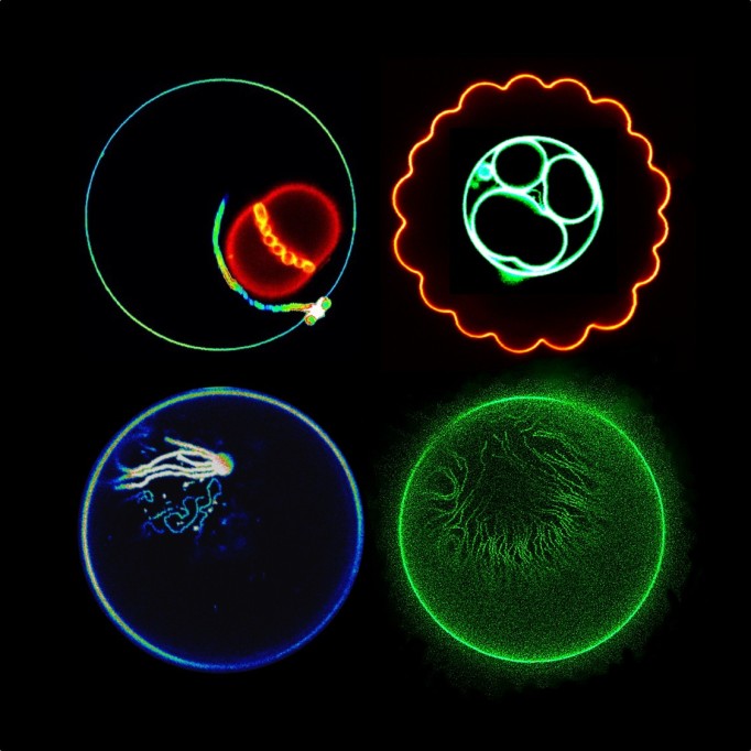
This final image actually consists of six individual images, each of them represents the various remarkable shape transformations of giant unilamellar vesicles (GUVs). These are cell-sized lipid vesicles in which the membrane can be directly observed under the microscope and tell you how it reacts to various substances and perturbations. The GUVs here encapsulate aqueous two-phase system (ATPS) of dextran and PEG solution (see e.g. http://dx.doi.org/10.1002/admi.201600451), which mimics the crowded environment in the cytosol. In practice, GUVs encapsulating ATPS represent an oversimplified version of the cell. Mimicking certain cell functionalities as is the case here, is one of the targets of the MaxSynBio consortium (https://www.maxsynbio.mpg.de/), which funds my work. When deflated, these ATPS-GUVs deform into all sorts of curious shapes. Some produce nanotubes protruding into the vesicle interior and accumulating at the ATPS interface. Others offer fascinating images like “the crying face” and “the jelly fish”, and this is what I call the beauty of science: sometimes doing nothing is better than doing something. This series of images was captured using confocal and STED (super resolution) microscopy, and assembled into a mini story: “The little fella is worried that his friends (snail and jellyfish) are being sucked into ‘the green hole.’”
What do you love about this image? Or, what about this image made you submit it for the contest?
I love the final combination of the image which enables storytelling. From the individual images, I like “the crying face” in particular. The moment I saw it during the experiment, two things flashed into my mind, Doraemon and the famous painting called “The Scream” by Edvard Munch. This image actually represents the beauty created by nature in my experiment; it cannot be controlled or replicated. When I first saw it, I already had the thought of combining it with other interesting images from my work into some funny picture. The good thing about doing microscopy work is that you get expected and unexpected results, and the images from both can astonish, even surprise and amaze you. The best time of the day is when I get some very expressive and interesting images from the experiment and share them with my girlfriend and my family ASAP. They are often amazed by how the micro-world in some way resembles the real-world. Fortunately, the Biophysical Society has this image contest at the Annual Meeting which provided me with the opportunity to show my work and also a little bit of this artistic image to the whole community.
What do you want viewers to see/think when they view this image?
I want to use my image to remind the viewers that beautiful things are everywhere. Please, always try to keep your mind open and see things from different perspectives. Even the “garbage” data for your experiments can be pretty and expressive in other ways. So, enjoy doing your work and it might surprise you in some other ways! Like winning an image contest 🙂
How does this image reflect your scientific research?
Internal structures in GUVs can respond to deflation in quite a curious way. The jellyfish structure in the image is presumably a smaller deflated pancake-like vesicle with protrusions swimming along the interface of the ATPS, which is a curved quasi two-dimensional surface. Such structures would be normally viewed as data to discard as the structure is difficult to resolve and we cannot conclude much about the membrane response. However, the image is curious and pleasing to the eye. Vesicles without initial internal structures are the most suitable for the project that I have been working on. Upon deflation, the excess membrane area of the GUV is stored in the form of membrane nanotubes protruding into the vesicle interior, and based on the composition of the lipid membrane, the shape of the nanotubes can be either necklace-like or cylindrical. The thickness can be below or above the optical resolution. My research focuses on investigating nanotubes like those in “the green hole” image. Their thickness is below the optical resolution, which is why I employ super resolution microscopy (STED) to resolve them. The diameter of these nanotubes reflects changes in the spontaneous (preferred) curvature of the membrane. The related article is in preparation and soon to be published, so stay tuned!
Can you please provide a few real-world examples of your research?
The nanotube formation occurs in GUVs exhibiting phase separation (i.e. droplet formation) in their interior crowded by macromolecules. In real life, phase separation in the lens cell can cause cataract which is a disease affecting countless number of people. Phase separation or droplet formation in cells is an emerging topic, which was also covered in a symposium within this annual meeting. Our studies with GUVs aim at answering the mechanism of cellular events and diseases associated with the contact and interaction of the droplets with the membrane. Our vesicles are loaded with high concentration of macromolecules. This solution can dynamically phase separate above a certain concertation into two droplets within the GUV. Thus, this system can certainly act as a more robust mimetic cell to interpret the much more complex cellular phenomena. So, studying how biomembranes restructure themselves in the environment of macromolecules can certainly promote our understanding on certain diseases in the human body and might even provide a future in fighting against them.
How does your research apply to those who are not working in your specific field?
Membrane nanotubes are ubiquitous and stand for the highly curved membrane structures and organelles in cells such as the endoplasmic reticulum and Golgi apparatus, as well as membrane vesicles generated for transport in and between the cells. They play a vital role in cellular events to regulate our life activities, and many of the mechanisms are still beyond our knowledge. Studying the generation and stabilization of their large curvature should be of immense importance to researchers in different fields.
Do you have a website where our readers can view your recent research?
More work on the nanotube formation in the ATPS GUVs can be found via these links, https://www.dimova.de/, and https://www.researchgate.net/profile/Ziliang_Zhao3.
I would like to acknowledge two people who helped me with the final version of this image. My girlfriend Yubing Guo (currently master student at Ruhr-University, Bochum) provided the idea of the snail and combined the snail image (top left corner) for me; my supervisor Dr. Rumiana Dimova removed the exterior signal of “the green hole” (bottom right corner) to make it “real,” provided the image with a beautiful name (Reshaping Vesicles via Deflation) and revised the wording in the description for me. I also acknowledge MaxSynBio and the head of our department, Prof. Lipowsky, for the funding and support.
